Ultrasound software
Open-source analysis software
fUSI research is booming, and there is an increasing demand for a standardized analysis workflow. The Urban Lab at NERF is developing the fUSI Analyzer (FSA), an open-access analytics pipeline for fUSI data analysis, in collaboration with many neurosciences and computer science teams.
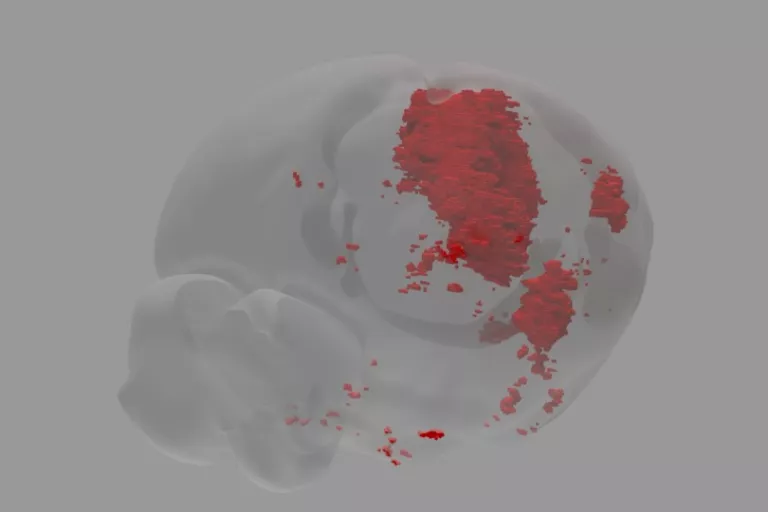
We are developing a dedicated software suite that provides a user-friendly graphical interface that allows users to analyze and display the fUSI data set. This software includes pre-processing functions like filtering, smoothing, and registration of the brain data to the mouse Allen Brain Atlas. It also provides a set of data analysis strategies such as correlation analysis, T-test, general linear model, spatiotemporal clustering, and resting-state analysis. Finally, the software improves data visualization, including several displaying functions to generate the region-time trace (barcode-like figure), the time course plot, the activation brain map, and the 3D rendering of the brain activation.
Several predefined analysis pipelines simplify data analysis for beginners and non-programmers based on standardized workflow. This software suite is a centerpiece of the fUSI ecosystem and may support a significant adoption of the fUSI technology among the scientific imaging community. The software is currently in beta testing and has already been distributed to several laboratories with a Creative Common open-source license. Ultimately, it will be made available through the standard channels (i.e., Github).
Automated brain registration and segmentation
Image registration is a crucial step in various biomedical imaging applications. It provides the ability to geometrically align one dataset with another and is a prerequisite for all imaging applications that compare datasets across subjects, imaging modalities, or time. Image segmentation is also essential in medical image analysis and is often the first and most critical step in many clinical applications. In fUSI analysis, image segmentation is commonly used for measuring and visualizing the brain's anatomical structures, analyzing brain changes, delineating pathological regions, and for surgical planning and image-guided interventions.
Registration and segmentation are challenging for a non-expert. It is both time-consuming and error-prone, which impairs data analytics and reproducibility. We are developing advanced solutions for geometrically transforming any acquired data (plane or volume) to match a reference coordinates framework on an atlas with high precision and in real time.
Digitization of vascular data
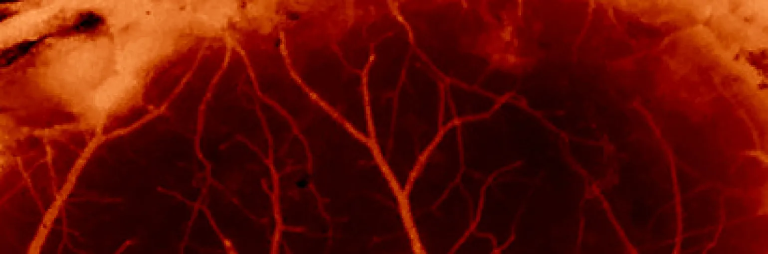
One way to assess the risk of cerebrovascular pathologies is to use computational models to predict the physiological effects of a reduction of blood supply and correlate these responses with observations of brain damage. It is, therefore, crucial to establish a detailed 3D organization of the brain vasculature. The Urban lab is currently developing an automated image segmentation and registration tool in collaboration with the IPI department, Dr. Danilo Babin, Dr. Bart Goosens and Dr. Wielfried Philips in Ghent, Belgium. This new automated tool based on deep learning is designed for real-time decision support.
High resolution imaging without contrast agent
We are developing new methods and algorithms to improve image quality, resolution and sensitivity.